C3.6 - Human Digestive Tract
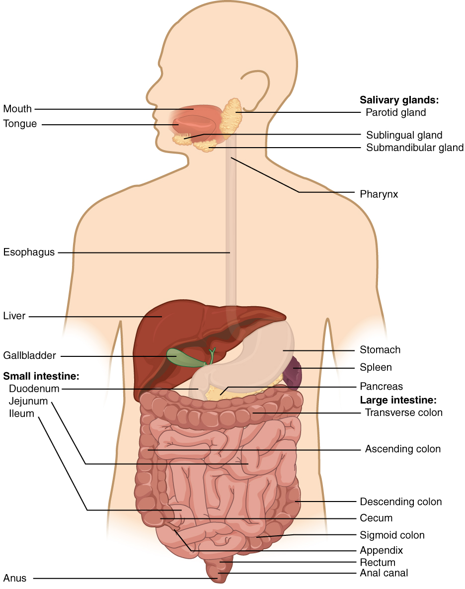
Diagram of Human Digestive Tract
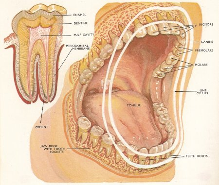
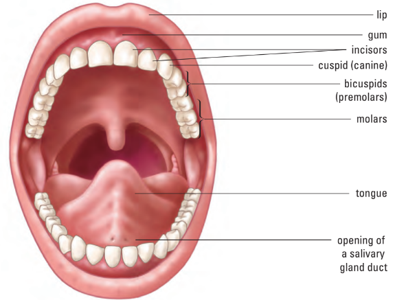
The Mouth and Dentition
Teeth
Mouth, easier to understand
-
Digestion begins in mouth
-
Food is broken into smaller pieces by teeth
-
Teeth
- 4 incisors
- 2 canines
- 4 bicuspids
- 6 molars on lower jaw
-
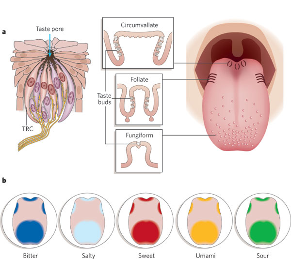
Tongue covered w/ papillae which house the taste buds
-
Taste buds detect
- sweet
- sour
- salty
- umami
- bitter
-
Roof of mouth divided into hard and soft palate
-
Uvula hangs from back edge of soft palate and prevents food from entering pharynx when swallowing
-
Salivary glands secrete saliva that contains enzymes such as amylase that start chemical digestive process.
-
saliva: water liquid that contains digestive enzymes, mucus, and other chemicals to help chemically digest food
-
mucus: sticky substance that coats and lubricates food to slide down tube without harming lining tissues
Pharynx
- After leaving mouth, bolus enters pharynx
- bolus: food ball
- pharynx: short tube shared by digestive and respiratory system
- epiglottis: cartilage flap that temporarily seals airway to prevent food from entering airway
Esophagus
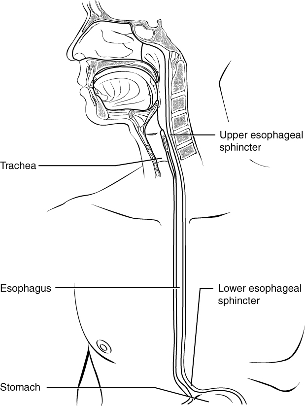
- After leaving pharynx, bolus enters esophagus
- esophagus: muscle-lined tube connecting the mouth and stomach
- Lubricant, mucin is secreted to aid in swallowing
- regurgitation: food returning up esophagus
Peristalsis
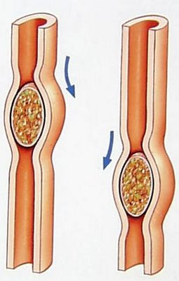
peristalsis: movement of food through wave-like contractions of muscle in digestive system
Stomach
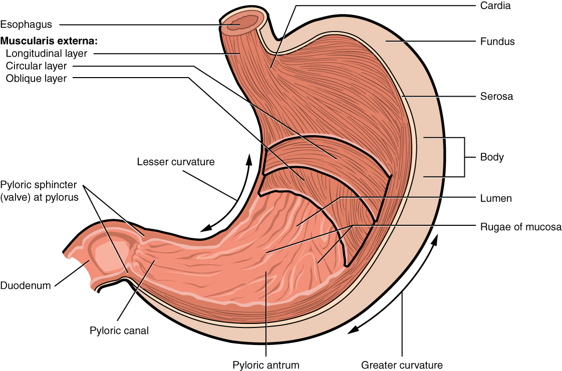
- stomach: elastic muscular, J-shaped sac that helps digest bolus
- Interior lined w/ 1000s of gastric juice secreting glands contained in gastric pits
- gastric juice: mixture of hydrochloric acid, enzymes like pepsinogen, and mucus
- Gastric juice and muscular movements render food into thick liquid called chyme
- chyme: acidic, nutrient-rich liquid produced by stomach
- Amount of food entering and leaving stomach controlled by 2 sphincters:
- esophagal -OR-
- cardiac + pyloric
- Stomach muscles contract, create churning motion to force chyme into small intest.
- pyloric sphincter: junction where stomach connects to duodenum (small intest.)
The Liver
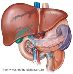
- Large organ, numerous roles
- hepato-: related to liver
- bile: yellow / greenish alkaline liquid produced by liver
- bile duct: passageway for bile to travel through
- Produce bile salts (alkaline organic salts) from cholesterol
- Bile emulsified or breaks up fats and activates lipases in small intestine
- Bile stored in gallbladder
- Breaks down old red blood cells producing bile pigments:
- biliverdin and bilirubin
- Detoxifies the blood
- Converts monosaccharides (animal starch) into glycogen
- Stores vitamins A, D, E, K
The Pancreas
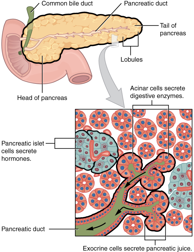
- Produces pancreatic juice
- pancreatic juice: clear alkaline liquid that neutralizes acidic chyme and contains enzymes to further break down nutrients
- pancreatic duct: passageway for pancreatic juices to pass through
- Produces enzymes such as pancreatic amylase and lipase
- Releases bicarbonate to neutralize acidic chyme in the duodenum
- Frederick Banting found way to preserve pancreatic insulin
- to treat Type I Diabetes
Diabetes
-
Diabetes – A group of diseases characterized by high blood glucose levels due to issues with insulin secretion, insulin action, or both.
-
Insulin – A hormone produced by the pancreas that controls blood glucose levels; discovered by Frederick Banting and Charles Best in 1921.
-
Type 1 Diabetes – An autoimmune condition where the pancreas cannot produce insulin; cause is not fully understood and currently not preventable.
-
Type 2 Diabetes – A metabolic disorder where body cells do not respond properly to insulin; strongly linked to obesity and lack of physical activity.
-
Effects of Diabetes – Includes fatigue, vision loss, kidney disease, heart problems, and high blood pressure due to persistent high blood glucose.
-
Lifestyle Impact – Sedentary habits and excess body fat hinder insulin effectiveness, increasing the risk of developing Type 2 diabetes.
The Small Intestines
- small intestine: tube w/ folded walls in the body
- villi: folds in the small intestine that increase surface area for absorption of nutrients
- Intestinal glands found within villi
- Villi have finger like projections called microvilli
- Gross cylindrical surface of 0-4 m2 (7-8 pgs. of textbook), incl. villi and microvilli
- Surface area is 300 m2 (doubles tennis court)
- Each villus has network of microscopic blood vessels and a small lymph vessel
- lymph vessel: structures that carry fluid away from cells
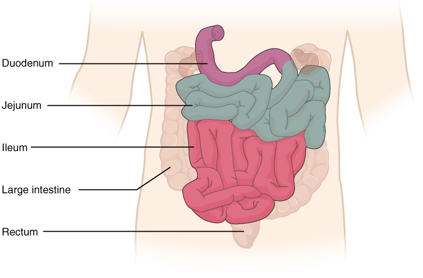
- Subdivided into 3 regions:
- duodenum: fairly short, U-shaped region connecting stomach to small intestine
- pancreatic and bile ducts open into duodenum
- jejunum: 2.5 m long region that contains more folds and intestinal glands than duodenum
- ileum: 3 m long region that contains fewer and smaller villi which absorbs nutrients broken down in duodenum and jejunum
- duodenum: fairly short, U-shaped region connecting stomach to small intestine
Large Intestine
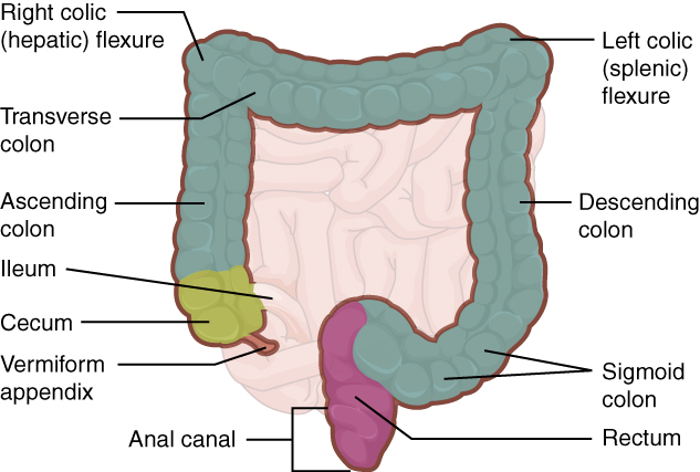
- large intestine: 1.5 m long large tube — subdivided into 4 regions:
- caecum: blind sack, appendix at extreme end
- colon: the main parts of the large intestine that absorbs water and minerals
- site of bacterial fermentation
- Food enters…
- ascending colon
- transverse colon
- descending colon
- Feces passes out of colon to rectum, then anal canal and sphincter
- feces: undigested food material and other waste products
Chemical Digestion
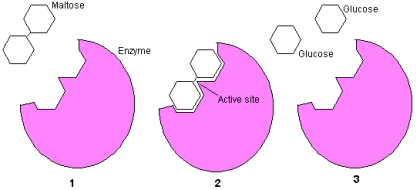
- digestion: chemical breakdown of food by hydrolysis (adding water molecule)
- Hydrolysis sped up by enzymes:
- carbohydrases (breaks down carbohydrates)
- lipases (breaks downfats)
- proteinases (breaks down proteins)
- Excreted from glands into digestive tract
- Major enzymes involved in digestion:
- salivary amylase (amylose)
- pepsin (protein)
- lipase (fats)
- maltase (maltose)
- lactase (lactose)
- Digestive secretions regulated by nerves and hormones
- hormones: chemical messengers transported from one part of the body to another through the blood
Diagnosing Digestive Disorders
Diagnostic Tools
- endoscopy: process where narrow tube inserted into throat and passed into esophagus, stomach, and upper intestine
- small camera in narrow tube allows doctor to see inside and spot problems
- biopsy: cutting of small piece of digestive tissue to view under microscope
- colonoscopy: thin, lighted tube w/ camera inserted through rectum to detect unusual growth
- capsule endoscope: pill with camera swallowed that takes picture of tract before being eliminated
Examples of Digestive Disorders
| Condition | Description | Diagnosis and Treatment |
|---|---|---|
| Gastroesophageal reflux disease (GERD) | Acid from the stomach frequently flows back into the esophagus. | Avoid smoking, alcohol, and overeating. Use antacids. Surgery may be needed in some cases. |
| Ulcer | Damage to stomach lining causes irritation by acid. | Tests like blood, stool, X-ray, and endoscopy identify the ulcer and its cause (often H. pylori). |
| Inflammatory Bowel Disease | Intestines become inflamed and develop ulcers. | Believed to be an allergic reaction to foreign substances in the intestines. Treated with medication or surgery. |
| Constipation | Stool moves too slowly or becomes too hard due to lack of fiber or other causes. | Eat a high-fiber diet, drink water, and exercise regularly. |
| Diarrhea | Colon is irritated, reducing water absorption, often due to infection. | Usually resolves with fluids and nutrition. May be a symptom of a more serious condition. |