C3.9 - Mammalian Circulatory System
Introduction
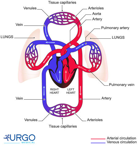
- Blood arranged in 3 main cycles below:
- cardiac circulation: pathway of blood within heart
- pulmonary circulation: pathway of blood from heart to lungs
- systemic circulation: pathway of blood from heart to rest of body
Transport Vessels
- Arteries and arterioles (small arteries) carry blood away from heart
- oxygenated blood
- pulmonary arteries carry deoxygenated blood to lungs
- Veins and venules (small veins) carry blood toward heart
- deoxygenated blood
- pulmonary veins carry oxygenated blood to heart
- blood vessel: a tube for blood to circulate through
- Pulmonary cycle breaks oxygenated-deoxygenated trend
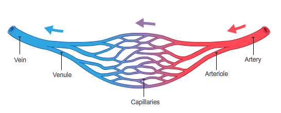
- Blood travels from arteries → capillaries → veins
- As blood travels through capillaries, pressure drops
Arteries
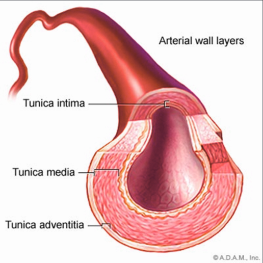
- arteries: thick, elastic, muscular transport vessels that carry blood away from heart
- Act as conduits for blood between heart and capillaries
- Act as a pressure reservoir to force blood into arterioles
- Dampens changes in blood pressure and flow caused by the heart
- Controls distribution of blood to different capillary networks by closing some arteries
- Walls of arteries made of epithelial tissue wrapped in layers of smooth muscle and connective tissue
- Blood pressure depends on…
- the volume of blood in the arteries
- properties of arterial walls
Veins
- veins: transport vessels that carry blood towards heart
- Large inside diamater
- Lack elasticity
- Large volume, low pressure systems
- Storage reservoir for blood
- Blood moves back to heart by gravity and skeletal muscle contractions
- exercise is important
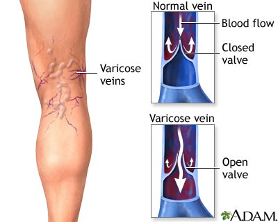
- Valves in veins prevent blood from flowing backwards
- Varicose veins: a disease where the valves in the veins do not function properly
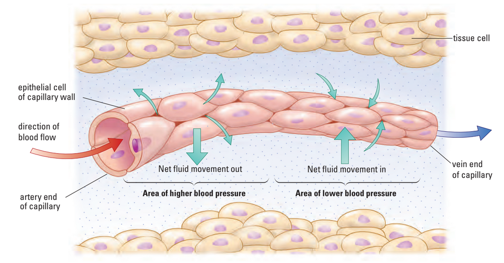
Capillaries
Line 1 blood cell thick are capillaries
- capillaries: smallest (microscopic) blood vessels where nutrient and gas exchange between blood and body occurs
- Diameter just large enough for 1 blood cell to pass through
- High surface area, resembles network of tiny tubes
- Capillary wall regulates movement of fluids and nutrients into and out of blood
- Walls consist of very thin layer of epithelial tissue in moist membrane
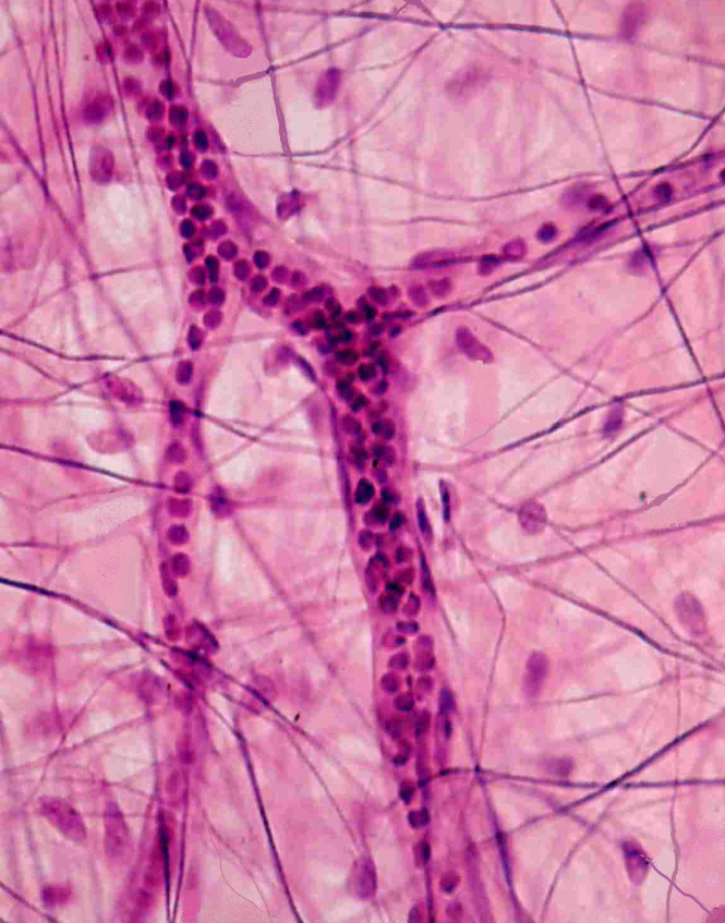
Blood: The Transport Medium
- tissue: collection of cells that have specialized tasks
- blood: transport tissue of oxygen and nutrients
- Body contains ~4-6 L of blood
- also transports wastes out of system
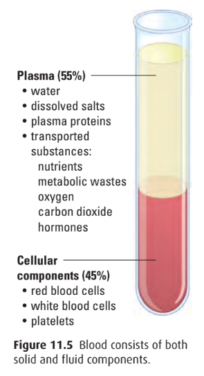
- Composed of 2 phases
- plasma: liquid part of blood (55% of blood)
- ~90% water
- ~10% of plasma: proteins, dissolved nutrients, wastes
- solid cells (45% of blood)
- plasma: liquid part of blood (55% of blood)
Erythrocytes: Red Blood Cells (RBCs)
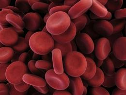
- red blood cells (RBCs): cells that carry oxygen from lungs to all tissues of body
- 1 mm3 of blood (1 drop) ≈ 5 mil. RBCs
- proper name: erythrocytes
- 44% of blood volume
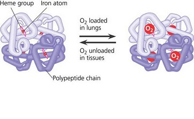
- Bags of hemoglobin, made in bone marrow
- hemoglobin: protein that binds oxygen in the lungs and releases it throughout body
- Each cell packed w/ 280M haemoglobin molecules
- Haemoglobin takes up and releases oxygen
- Oxygen bonds to Fe in hemoglobin, giving blood red colour
- Specialized for oxygen transport
- Produced in bone marrow
- Mature cells have no nuclei
- Lifespan: 3-4 mo.
- Factors that influence oxygen-haemoglobin binding
- Low oxygen partial pressure: weakened bond, quick release
- Increased blood acidity: weakened bond, quick release
- Cooler temperature: strengthened bond, slow release
- Carbon dioxide also binds w/ haemoglobin
Haemoglobin binding diagram
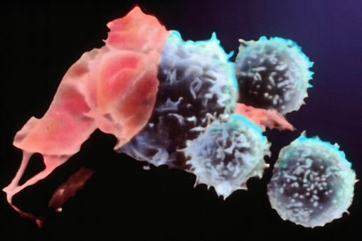
Leukocytes: White Blood Cells (WBCs)
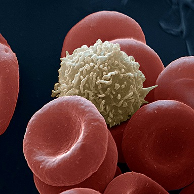
- white blood cells (WBCs): cells that guard against infection, fight parasites, and attack bacteria
- proper name: leukocytes
- 1% of total blood volume under normal, healthy conditions
- Protect body from infection
- Not constrained to blood vessels, can move through vessel walls as needed
- Keep their nuclei unlike RBCs
- 2 important types
- macrophages
- lymphocytes
- Macrophages may pass through capillary wall by amoeboid movements
- to digest pathogens by phagocytosis
- Part of innate immune response: general defence
Lymphocytes
- Lymphocytes part of acquired immune response
- Enable body to recognize and fend off pathogens
- 2 types
- T cells (from thymus gland)
- B cells (from bone marrow)
- Allow body to become immune to certain pathogens
- antibodies: Y-shaped proteins w/ a region that is variable in structure
- Variable region recognizes antigen carried on invading pathogens
- B cells remain in blood after an infection ready to trigger another immune response
Platelets
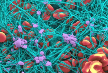
- platelets: cell fragments involved in clotting of blood
- originate when cytoplasm of certain bone marrow cells divides
- 1 mm3 of blood ≈ 250k-500k platelets
- Platelets mainly produced in bone marrow
- After injury, broken blood vessels attract platelets to site of injury
- Platelets rupture and release substances that combine with other clotting factors in plasma
- Enzymatic reactions produce fibrin that forms a mesh preventing blood cells from escaping
- fibrin: strand-like protein involved in clotting
- Network builds up into a scab
- Protects area while new tissue develops
Nutrient Exchange Between Blood and Cells
- diffusion: process where molecules move across a membrane from area of high concentration to low conc.
- diffusion gradient: gradual change in concentration of solutes in a solution as a func. of dist. through solution
- i.e. if blood in capillary contains higher conc. of O2 than fluid next to it…
- … oxygen moves to fluid by diffusion (spont.)
- Capillaries no more farther than 10μm from body cell
- Nutrients, gases, and wastes diffuse between capillaries and body cells
- Body tissues are surrounded by fluid that acts as a medium for molecular exchange.
- Molecules in capillaries must first enter the surrounding fluid before entering cells
- Small molecules (e.g., oxygen and carbon dioxide) move by diffusion:
- Oxygen diffuses from blood → fluid → cells
- Carbon dioxide diffuses from cells → fluid → blood
- Larger molecules require other transport processes to cross membranes
- blood pressure: force exerted by blood on artery walls which drives:
- Blood flow through arteries and capillaries
- Movement of substances across capillary walls
- Nutrients and gases diffuse into cells from the fluid.
- Cellular wastes diffuse out of cells into the fluid and then into the blood.
- Blood transports wastes to organs for excretion from the body.